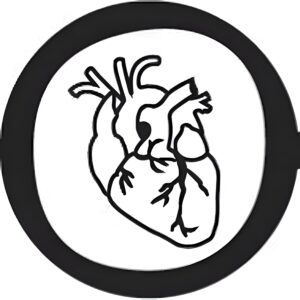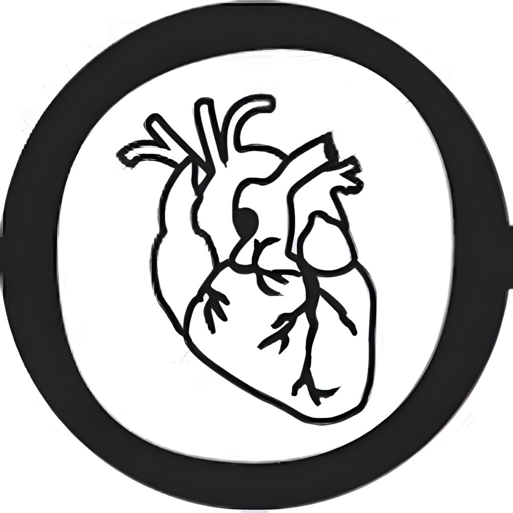
Glands, Tissue Healing, Cell-Cell Junctions, & Skin (5)
- September 16, 2025
- 12:08 pm
This video explores Week 5 content from Chapter 4 in detail.
It discusses the process of tissue healing and the role of glandular epithelium.
The structure and function of various tissues are explained with examples.
It also highlights different types of cell-to-cell junctions and their importance.
Lastly, the video includes a short labeling exercise on the layers of the skin.
Summary
Reviewed endocrine vs. exocrine glands and their secretions, cell-to-cell connections (tight junctions, adherens, desmosomes, gap junctions, hemidesmosomes), tissue healing processes (macrophages, fibroblasts, chemotactic agents like histamines, prostaglandins, leukotrienes), skin structure (epidermis, dermis, subcutaneous), epidermal layers, and burn classifications (first to third degree).
Raw Transcript
[00:00] Alright y'all, you ready? Week 5, let's go. We're going to review a few different things. We kind of hopped around a little bit. So this is what we're going to be going through. We're going to be going through the glans, endocrine versus exocrine, how they're different, how they're similar. We're also going to talk about the structure that helps
[00:20] its function as always. We're going to talk a little bit about cell to cell connections. I'm going to use a sheet that I handed out in class. We're going to talk a little bit about tissue healing and how we recruit different things to come to the site of damage to help it heal. Then lastly we're going to talk about skin layers and burn.
[00:40] and how they relate to the skin layer. So let's get started. First off, glands. Glans are basically collections of epithelial cells. So these are epithelial cells that secrete some form of fluid or another. Now in the name we can assume what's going to be the first part of the process.
[01:00] going on here. So for exocrine, exo means outside and crin literally means either to cry in Latin, but I'm going to say to secrete. So it's basically crying tears of secretions, crying tears of secretions.
[01:20] So in exocrine glands, the secretions that are made by these cells, these secretory cells, are eventually going to make their way out of the body. Okay, so let me show you a gland like that. This is one type of gland. There are several different orientations. But the biggest thing that you can see with exocrine glands especially is that they are very long-
[01:40] and branched. This allows for increased surface area on the apical side, these top sides of the cells, to basically allow for more cells to pack into one space to increase the amount of secretions that they can make. Now these are epithelial tissues. Like I said, they are usually cuboidal.
[02:00] epithelial cells or coloneus. The reason being is because these are pretty thick, big cells that have room for organelles to actually make the secretions themselves, that you need organelles to make them. Second. So like I was saying, cuboidal and coloneus have more space.
[02:20] space for organelles to make those secretions and that's why they're bigger. If they were squamous cells, if they were all flat like this, there's like no room for organelles, there's no room for stuff to go on in those cells. So that's why they are columnar organelles. In this case it's going to be cuboidal. When they secrete their fluid, they're going to secrete it into
[02:40] large opening, well large in terms of their size, into their larger opening called the lumen. Lumen is just a name for a hollow tube or hole. So you have gut lumen, that's like the inside of the gut, that hollow tube, where all of these secretory cells are going to secrete their fluid into the lumen and eventually go out to the lumen.
[03:00] out via a duct. That's the key indicator that it is an exocrine gland. It contains a duct that will then go out of the body. For example, we've got several different glands that have ducts. You've got your salivary glands that will eventually go into the hollow tube of your mouth.
[03:20] so technically out of your body. You can swallow, but then the swallowing, it would go out of your body eventually. Salivary glands would make saliva, so that would be the secretions. You could have sweat glands. So sweat glands, they would secrete sweat into that area, go out with the duct, get on your skin, cool you off.
[03:40] We also talked about, I'm blanking for a second, give me a second, sweat glands, sebaceous glands. These are very active during adolescence because you are secreting a lot of oils and that's why you get acme sometimes, these overactive sebaceous glands. And then you can also think of teary.
[04:00] ducts. Tear ducts they generate tears and it comes out of your eyes and your tear ducts. The other one I'm going to tell you about is the pancreatic fluid. So pancreatic fluid contains sodium bicarbonate. This is an
[04:20] axocrin gland because the sodium bicarbonate, as we talked about in previous videos, goes into your small intestine, your first part of it is the duodenum, and it's going to go in there and eventually make its way out of the body. But the reason it's secreting into that hollow opening is because it's neutralizing all the acid from your stomach. So since it is going out of the body,
[04:40] eventually it is considered exocrine and that will have a duct. We'll see the pancreas again in the endocrine system though too. So that is the exocrine glands, their shape, their function, and some examples. Awesome. Endocrine. Endo means inside and when we're talking about
[05:00] endocrine glands, they're going to be inside secreting into the blood. So that's the key for endocrine. Something's getting secreted directly into the bloodstream, as opposed to a duct going outside of the body. It is an endocrine function. It's an endocrine gland or an endocrine cell.
[05:20] So for these examples, we've got our cells. Say this is in your pituitary gland, so that would be a great example of an endocumulan pituitary. It's at the base of your skull. And it's going to secrete different secretions into the bloodstream. It usually does this by exosyptase.
[05:40] or just by simple diffusion, but we'll say exocytosis, you can remember it. Remember the vesicles will be surrounding it, it'll eventually fuse with the membrane and now it will get in the interstitial space, eventually get into the bloodstream. So all of these are usually point hormones because hormones
[06:00] are basically chemical messengers carried in the blood. So endocrine functions always go into the bloodstream via hormones and that is the secretions that those cells give out. So the big difference between exo and endo goes into the blood, exocrine goes out of the body through a duct.
[06:20] A couple other examples. The pituitary gland has a ton of hormones. The thyroid has a few but it's regulated by the pituitary. So the pituitary is like the master regulator. And then I wanted to tell you about the pancreas. Specifically pancreatic beta cells, these secrete
[06:40] insulin and if you could guess where does the insulin go? Into the bloodstream. So insulin is a hormone that goes into the bloodstream from the pancreatic beta cells because insulin is going to kind of act as a lock or sorry a key that opens the lock to the cells so it opens the gates up
[07:00] in the cells to allow glucose to flow in. So when you have high blood sugar, you just eat a high carbohydrate meal, your blood sugar is going to raise and you need all that glucose to get into your cells. So insulin is the little key that opens the lock, the ligand data channel, remember that, that allows glucose to flow into the cells, thereby that cell can now make an ETA.
[07:20] by breaking up the glucose. So that is exocrine versus endocrine. Endocrine glands, they have an entire system dedicated to it in anatomy and physiology too. I have a review video up for that if you'd like to watch it. Okay, so we got done with our glands and now let's talk about these cell-to-cell connections. It might not take too much time with these. You should have received
[07:40] this paper, you can also see it online. But there's several cell-to-cell adhesions that I want to talk to you about. First off, when we look at like a gland, like all these cells, it almost looks like they're sharing a membrane, but they're not. They usually are connected by some of these cell-to-cell connections. So imagine this.
[08:00] little section in the middle is in between each and every one of the cells to keep them together for certain tissue types. So let's walk through them. Top one, tight junctions. You see the plasma membrane is coming out a bit. Since the plasma membrane is hydrophobic, this is a water-tight junction.
[08:20] water cannot pass through these tight junctions very well, nor can many things at all. The adherence junctions in the desmosomes both connect them through these integral proteins in the membrane as well as these filaments that go inside the membrane themselves. So there's both an intracellular component and an extracellular component. The adherence-
[08:40] In other words, the difference, A, is connected by actin. This is a thinner filament, so it's more flexible, but it's less strong. Whereas the desmosomes, they have very thick, intermediate filaments, and they're much stronger and more stable, okay? Gap junctions as we move down
[09:00] They actually share, it's like a tunnel between cells. They share both a plasma membrane and cytoplasm. And this allows, this is what I want you to write down, ions to flow quickly from one cell to another. So if this cell were to gain a bunch of positive ions, well, normally cells are negative. So if this negative cell over here is
[09:20] connected by a gap junction to a positive cell now that has a lot of positive ions. Those positive ions are going to flow through the gap junctions into the other cell. This happens in your heart a lot through intercalated discs. You may have heard of those in lab with your cardiac muscle. Lastly you got hemidesmosomes, hemi meaning half, and
[09:40] Desmosomes usually connect a cell to a cell, but a hemidesmosome connects a cell to the extracellular matrix. And this is the hemidesmosome. This is an actin-linked one. It's not a desmosome, but I'm only going to talk about the hemidesmosomes on a test. I wouldn't talk to you about this one. So don't worry about that.
[10:00] types of cell to cell connections. That's basically all I went through in class. And there's several others. I was kind of grouping them into small categories for you. Alright, last couple things. Tissue healing. Look at my beautiful diagram over here. It's going to be hanging in an art show here very soon, sold for millions. Let's say you get a cut on your wrists.
[10:20] your carpal region. If you were to zoom in on that cut, you would see this opening that you just burst a bunch of cells open and potentially if it's bleeding you got into your dermal layer which we'll talk about the skin here in a second. So there's a blood supply so you start bleeding. Squeaky Wheel gets the grease. We want to fix the area that's broken.
[10:40] So we need to get some things to that area to heal it up. There are two things that I talked about that will go there. Macrophashes, big eaters to basically clean up the area, eat any bacteria or viruses that might get into there. And then fibroblasts, fibro meaning fiber, blast meaning to build. So it's going to build fibers.
[11:00] to basically plug up this hole that we've just made into our bloodstream to plug it up so that we don't bleed anymore. And that's also a platelet-mediated process, and we'll talk about that later on in A&P too. So we need to get these guys to that site. Right now they're not there. They're over here. We need them to move to that area.
[11:20] What do we use? Chemotaxic agents. So in that area, there will be a release of chemotaxic agents that will kind of slowly move out like this from high density to low density. And these chemotaxic agents I was drawn in this cover.
[11:40] chemo, emotaxic agents or molecules. Hemo means chemical, taxi means to move, right, be it taxied around. So it's basically chemicals that induce cells to move towards them. So the cells will detect these emotaxic
[12:00] molecules. And basically, just like climbing a ladder, they're going to follow those chemicals to the site of the breakage. It's a fascinating process. There's three main ones that I want you to remember about black numberings. There are three types. Histamines. You may have taken anti-histamines. They help with the inflammation.
[12:20] So histamines bump up inflammation, the redness so that you get a bunch of healing in one area. And antihistamines help when your nasal cavity specifically is inflamed from pollen, from allergens. And antihistamines kind of diminish that inflammatory response so the swelling goes down and you feel better.
[12:40] Those are a type of chemotaxic agent. Prostaglandins are another. Prostaglandins. And then lastly, leukotrins, leuko, meaning white. So leukotrins bring white blood cells to the area. And what's nice about your body is you'll
[13:00] have white blood cells kind of hanging out right underneath this epidermal layer so they're just ready to roll so they're ready to move where they need to go to fix the infection or to fight off the infection. I think that's all I went through in class so I'm going to stick it there. So I use this analogy. The chemotaxic agents are like the Batman.
[13:20] signal in the sky in Batman are the deposit fiberglass right wherever that is they're going to migrate to that area. Awesome. Last thing this was very short in my Monday Wednesday class you talked about the layers of skin. So if you look up here there's three distinct layers of
[13:40] skin. This is most superficial. This will be the epidermis, epi meaning on top of, therefore this section is the dermis. The epidermis is on top of the dermis and then this is the subcutaneous tissue. You may hear it be called the hypodermis. I like that.
[14:00] terminology because it's underneath the dermis. The epidermis is stratified squamous epithelial cells, so those flat layers of cells. The dermis contains like every type of tissue. You're going to have some simple cupoidal in your glands. You're going to have some
[14:20] dense irregular connective collagenous tissue. Under here, kind of keeping everything together, you're going to have variola right underneath the epidermis. There's a bunch, right? There's a bunch of tissues in the dermis. So this is a big plan right here. Subcutaneous is primarily adipose tissue, so fat.
[14:40] cells and that's going to connect to your muscles. Okay so it's all connected. There's also five layers of the epidermis. I'm going to label them from superficial to deep and they're all preface by stratum. So if I say stratum something that's one layer of.
[15:00] the epidermis because stratum means layer. So the most superficial layer, actually let me remind you, there's a great mnemonic, it's come let's get sunburnt. Come let's get sunburnt. So the first layer on the outside is the stratum cornea.
[15:20] The second layer is the stratum delucidum. Next one is rambulosum. Next one is spinosum. And the last one aptly named is the basale or the basal layer.
[15:40] all the way and get enough. So kind of makes sense, the corneum is the outside, corneas the outer part of your eyes. Lucidum is light, it's very clear, lucid dreaming is clear. Granulosum, there's granules. Spinosum, I hate because there's nothing specific about them, but it's right above the basal layer. And as
[16:00] As skin cells divide, so once again this is just in the epidermal layer, as skin cells divide in the basel, the stratum basel, they're going to push upwards. So all of these cells are going to get pushed upwards eventually. And if they get pushed upwards, uh-oh, they're getting away from the blood cell.
[16:20] supply that's in the dermis. If they get away from the blood supply, they'll lose their nutrients, therefore they will eventually die and slough off. They'll also become keratinized, which gives them a protein, a stronger barrier on the outside. So that would be keratinized, stratified squamous epithelial tissue. Lots of tissues here.
[16:40] Burns, last thing. You've got three types of burns. You got first degree, second degree, third degree. Third degree is the worst. First degree is I guess the best even though no burn is good. Epidermal layer. If it burns through, first degree burn. Through the dermis, second degree burn. Third degree burn.
[17:00] through the subcutaneous, so very deep. Either second and third degree burns both, require skin grafts, so basically putting skin on top of it, because you basically open up your body. So if you cut here, you got blood vessels in here. So now your body is open to the environment, ready for bacteria to just jump right into your bloodstream.
[17:20] So you need to cover that up with a skin graft in order to prevent infection. All right. Hey, that's week five, guys. Hope you learned a lot. Hope this was helpful. Drop any questions in the comments below, like and subscribe. Maybe I'll give you extra credit for it. We'll see. Take care.